Veterianary case study: It's not always a simple urinary tract infection

Sherman
Lyssa Alexander | Contributor
Sherman is a 6-year-old male Lhasa Apso. He is a very sweet dog who we been taking care of since he was a puppy. In early July, his owner brought him to the clinic for a problem we had never seen in him before. Every time he started to pee, he would strain and only produce a little bit of urine at a time. His owners also saw some blood in the urine, which prompted them to bring him in right away.
Sherman's physical exam was fairly normal. We collected a urine sample and took a look at it under the microscope.
A urinalysis is one of the most useful diagnostic tools that we have. With this simple, non-invasive test, we can see evidence of urinary tract infections, urinary tract tumors, kidney disease, metabolic disease, diabetes and much more.
In Sherman's case, there was pretty clear evidence of a urinary tract infection. Some urinary tract infections are uncomplicated. We put the dog on antibiotics and all the signs clear up rapidly.
However, sometimes there are additional factors present that predispose an animal to forming the infection in the first place. To check for these additional diseases, veterinarians need to perform bloodwork, a urine culture, radiographs (x-rays) and an ultrasound. This type of extensive workup is cost prohibitive in many cases, so we often have to select a smaller workup and do the best we can.

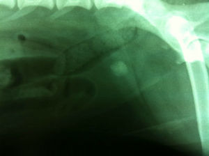
There is one stone in his bladder and a small stone inside the bone that runs through the urethra.
In Sherman's case, I was not satisfied with presuming that he had a simple urinary tract infection. There were several reasons for this. First was that he had never had urinary disease before.
The second was his breed. Lhasa Apsos are somewhat prone to stone formation. For these reasons, we decided to perform radiographs to investigate a little deeper. Sure enough, poor Sherman had two stones. One larger stone was located in his bladder and a second smaller stone was located in his urethra. Stones can cause a lot of irritation and harbor bacteria, leading to recurrent urinary tract infections. They can also grow and become obstructive, which can be a life-threatening condition. One way or another, they have to come out.

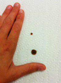
Sherman's calcium oxalate urethral stones.
Some stones can be dissolved with special diets, even if they are very large. Unfortunately, calcium oxalate stones do not dissolve.
To remove the stones, veterinarians can take them out surgically (cystotomy), express them manually under anesthesia (voiding urohydropropulsion), remove them with a tiny camera and special catheter (urethroscopy techniques), or break them apart with specialized equipment (electrohydraulic lithotripsy, laser lithotripsy). Based on the size of the stone, the cost of the procedures and the availability of specialized equipment, we elected to remove the stones surgically.

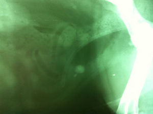
This image shows two stones in Sherman's bladder after successful retrograde urohydropropulsion.
Lyssa Alexander | Contributor
Therefore, we performed a procedure called retrograde urohydropropulsion. This involves using a catheter to flush the stone out of the urethra and back up into the bladder. Once all the stones are in the bladder, they can be retrieved with one surgery. Luckily, this procedure was successful, and the stone in his urethra moved back into the bladder easily. Once the stones were together, his bladder was opened surgically and the two stones were retrieved.

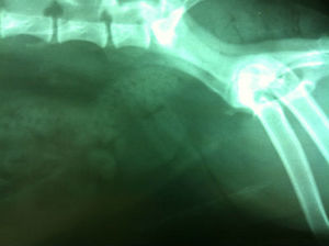
This image was taken just after Sherman's surgery to confirm that all his stones have been removed.
Lyssa Alexander | Contributor
Sherman is a prime example of how advanced diagnostics can help us to fully understand a case and pick out the best therapy. Thank you so much to Sherman's owners for sharing their story and for taking such great care of their pup!
Lyssa Alexander, DVM treats small and exotic animals and pocket pets at All Creatures Animal Clinic.

